Tiggy – Radius and Ulna fracture
Meet Tiggy
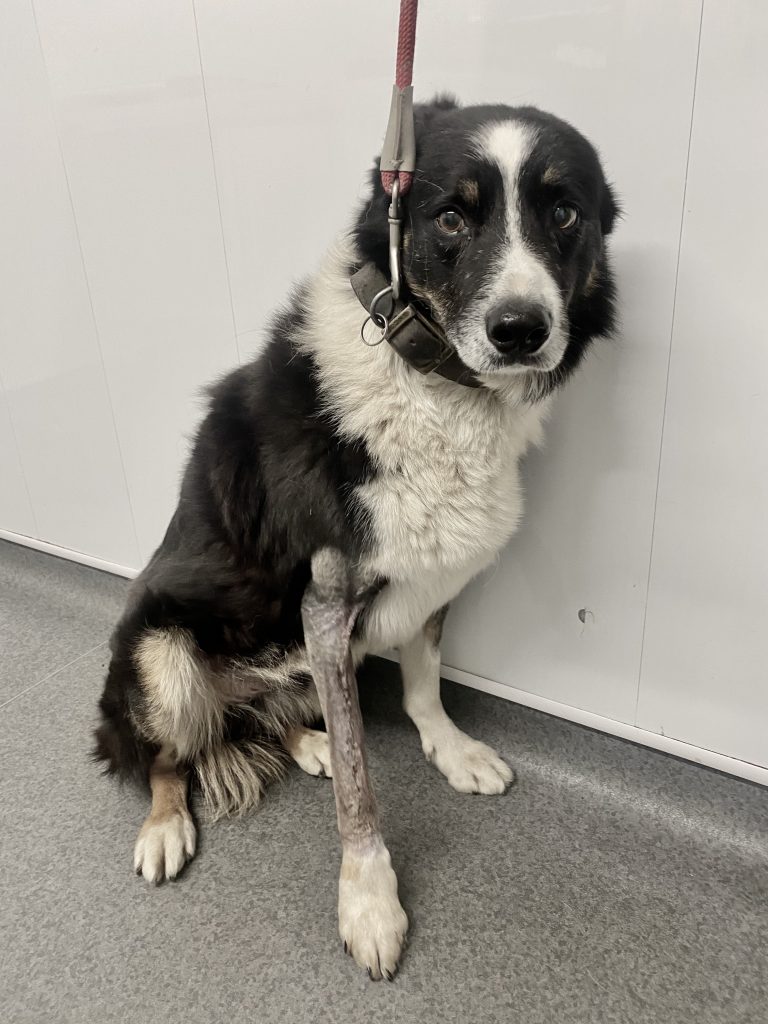
The Problem
Tiggy had sustained a complete, displaced fracture of the radius and ulna. With both bones broken, the limb was unstable and surgical stabilisation was recommended to restore alignment and allow the bones to heal. Your Goals with Our Expert Guidance
How do we describe a fracture?
Not all broken bones are the same. Classifying a fracture helps us describe exactly what has happened and plan the most appropriate method of repair.
We consider several features, including:
- Whether the skin remains intact (closed) or there is an associated wound communicating with the fracture (open).
- Which bone is affected and where along the bone the fracture has occurred.
- Whether the fracture is complete or incomplete.
- The shape of the fracture line – for example transverse, oblique, spiral or more complex with several fragments (comminuted). Nature/number of fragments.
- How much the pieces of bone have moved, shortened or rotated.
- Whether the fracture involves a joint or growth plate.
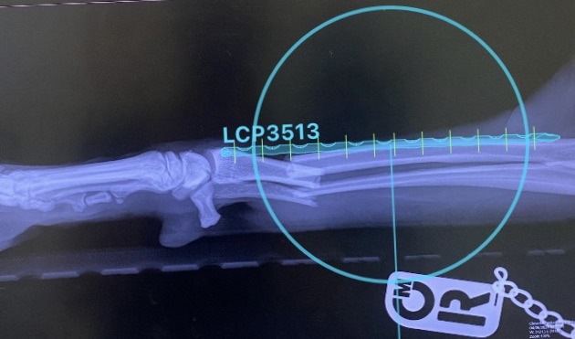
For Tiggy, his injury could therefore be described as a Closed, complete, transverse fractures of the distal diaphysis of the radius and ulna, with moderate caudal displacement of the distal fracture fragments.
Planning Tiggy’s repair
The plate is positioned over the radiograph before surgery to help assess the length and placement required.
Surgical Treatment
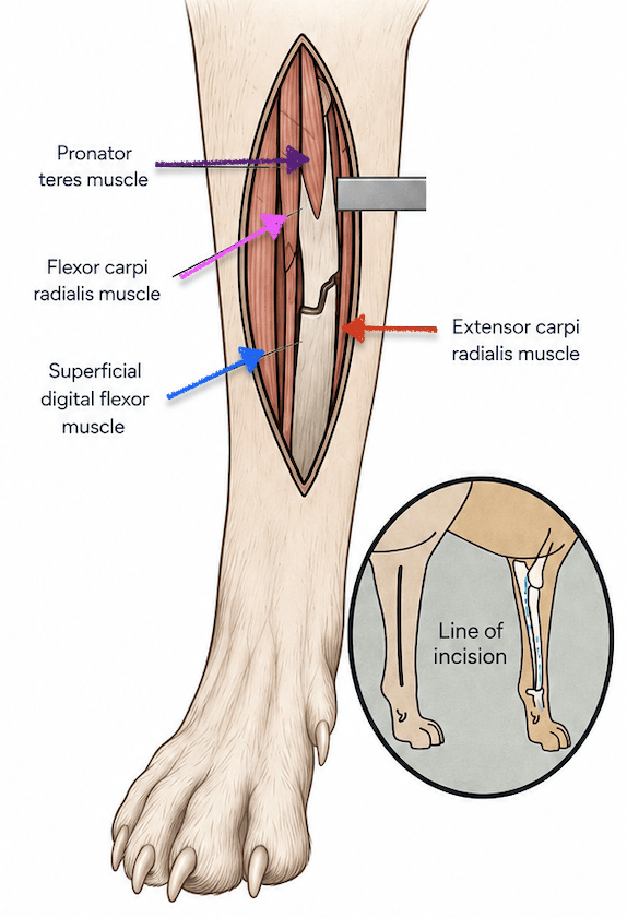
A craniomedial approach to the radius was made to expose the fracture while carefully preserving the surrounding soft tissues and blood supply. The fracture ends were identified, cleared of interposed tissue where necessary and carefully manipulated back into their normal anatomical alignment.
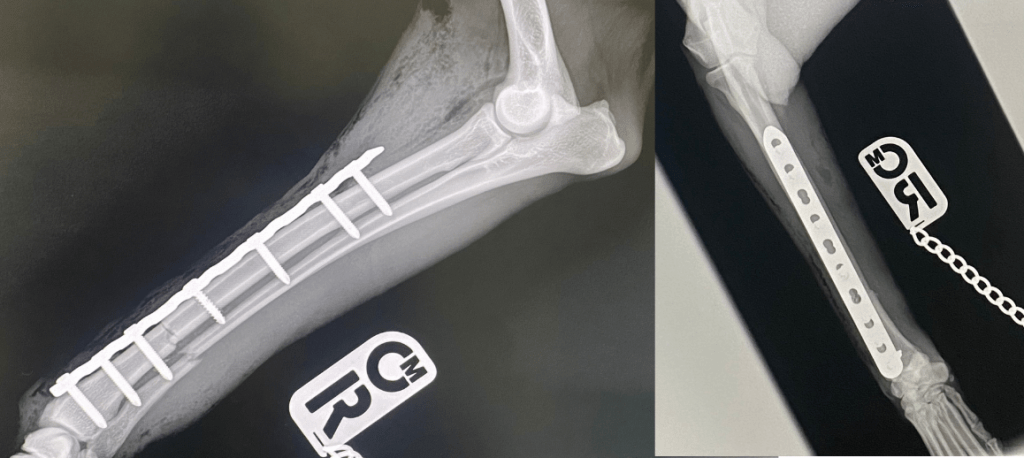
Once reduction had been achieved, the position, length and rotation of the radius were assessed before fixation. A long locking compression plate (LCP) plate was contoured to the craniomedial surface of the radius, spanning the fracture and allowing secure screw placement into the intact bone on either side.
The plate was applied to provide stable fixation and compression across the fracture, maintaining the restored length and alignment of the radius while healing took place. The associated ulnar fracture did not require separate fixation; stabilising the radius provided sufficient stability for the ulna to heal alongside it.
The surgical site was thoroughly lavaged before the soft tissues and skin were closed routinely.
Post-operative radiographs were then taken to assess the repair. These confirmed good reduction of the fracture, restoration of the normal alignment of the limb and satisfactory positioning of the plate and screws.
After the surgery
Post-operative radiographs were then taken to assess the repair. These confirmed good reduction of the fracture, restoration of the normal alignment of the limb and satisfactory positioning of the plate and screws.
Summary
For a working farm dog, the hardest part of Tiggy’s recovery may have been the prescription for strict rest and staying clean!
Exercise was carefully restricted while new bone formed around the fracture. Follow-up radiographs were used to assess healing before his activity was gradually increased.
Once sufficient healing had occurred, Tiggy was able to leave enforced rest behind and return to the fields he adores. 🐾
Advanced Surgical support for Veterinary Practices in Durham, Stockton on Tees, Northallerton, and Darlington.
Contact us to discuss your cases.